

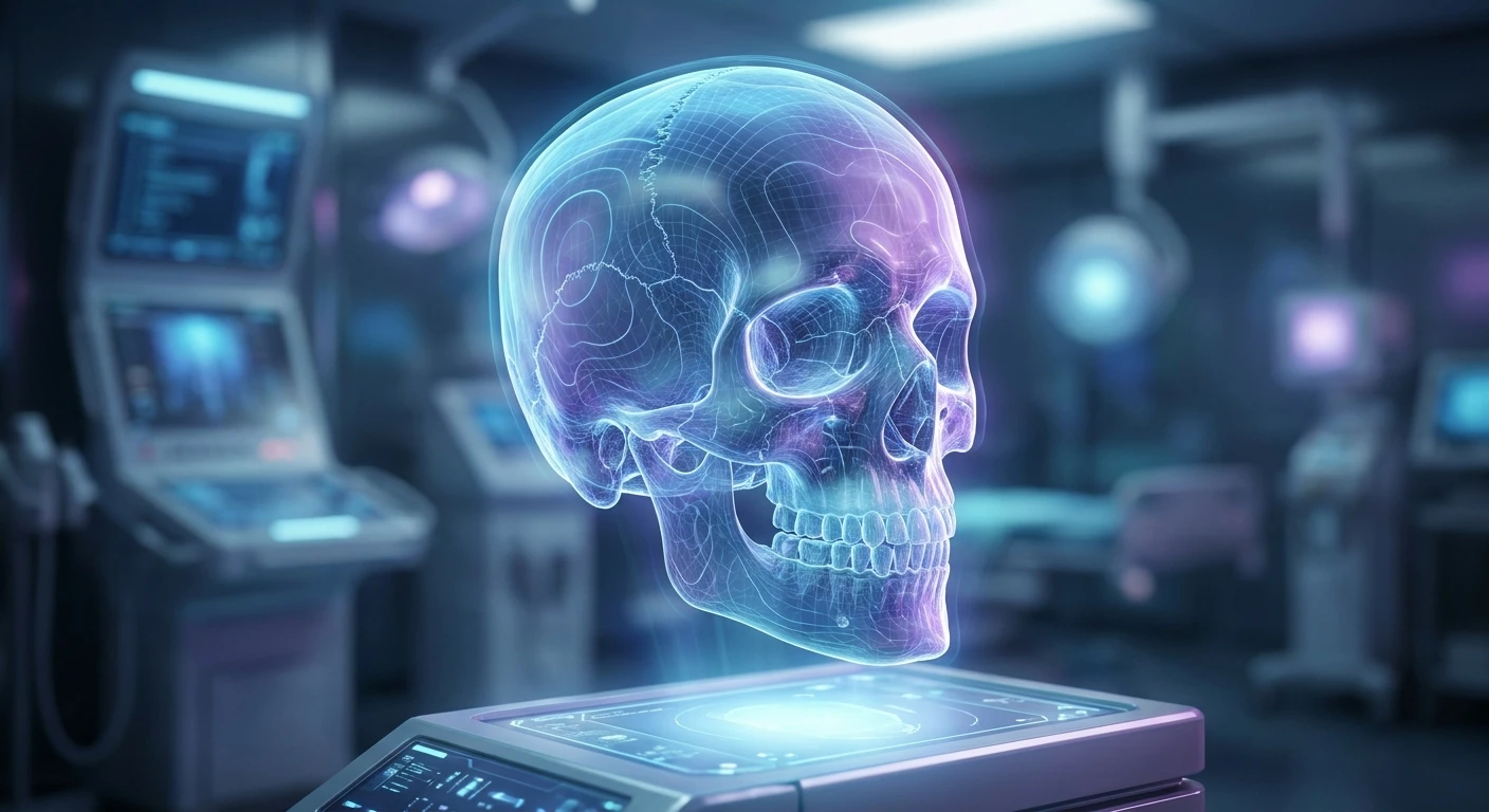
Understanding a Dent in Head: Causes and Concerns
Finding a dent in head suddenly can trigger immediate worry, sending your mind racing through the worst possible scenarios. The human skull is naturally bumpy and uneven, but feeling an unexpected depression or indentation warrants proper understanding rather than panic. The underlying truth is that most variations in cranial shape are completely benign and result from genetics, old minor injuries, or simple structural differences. However, determining whether an indentation requires medical attention means looking closely at the surrounding symptoms and the speed at which it appeared. This guide breaks down the biological facts about cranial depressions, separating harmless anatomical quirks from situations that need a doctor’s evaluation. We will walk through the exact structural makeup of the skull, the medical conditions linked to cranial changes, and actionable steps to evaluate your specific situation safely. Your skull acts as a rigid, protective helmet for the brain, consisting of multiple plates that fuse together during early development. That fusion process leaves behind natural ridges and valleys. While a dent in head is often just a reminder of childhood bumps or natural anatomy, grasping the mechanics behind it provides ultimate peace of mind. It empowers you to make informed health choices quickly and confidently without unnecessary stress. By addressing the root causes and recognizing when to act, you take control of your physical well-being.
When assessing a cranial indentation, you need a clear framework to distinguish harmless variations from medical emergencies. The core issue revolves around tissue compression, bone structure alterations, and potential trauma. A common example is wearing tight headphones or glasses, which can temporarily compress the soft tissue and scalp, creating the illusion of a bone dent. Another example is a depressed skull fracture resulting from a high-impact fall or blunt force trauma, where the bone physically caves inward, demanding emergency neurosurgical intervention.
Read also: Symptoms of Montgomery Gland Cancer Explained; Sudden Sharp Pain Under Left Breast Female: Causes.
| Indentation Type | Primary Cause | Typical Required Action |
|---|---|---|
| Soft Tissue Dent | Prolonged pressure from headwear | Remove pressure, wait for tissue rebound |
| Congenital Suture | Natural bone fusion at birth | None, completely harmless anomaly |
| Bone Trauma | Direct physical impact | Immediate medical evaluation and scanning |
If you notice a depression, follow this initial evaluation protocol to gather baseline information before consulting a physician:
- Assess for physical symptoms: Check for headaches, vision changes, confusion, nausea, or localized pain near the depressed area.
- Review recent activities: Think back over the past few weeks for any head injuries, falls, or use of tight headgear that could explain the change.
- Monitor the progression: Note whether the depression is expanding, changing shape, or becoming tender over time.
These steps form the foundation of a logical medical assessment. Your scalp contains layers of skin, fat, and muscle that can easily mimic structural bone issues when compressed. Understanding the distinction between soft tissue displacement and true skeletal depression prevents unnecessary anxiety while ensuring you act decisively when severe trauma actually occurs. Keep track of how the area feels to the touch—soft and pliable versus hard and rigid. Recognizing these nuances makes communication with a healthcare provider much more effective. Evaluating these factors objectively prevents panic while prioritizing genuine medical safety. You must remain vigilant about accompanying symptoms.
Origins of Skull Assessment
Humanity has observed and documented variations in skull shape since ancient times. Early physicians and healers frequently noted physical anomalies like a dent in head, often attributing them to spiritual or supernatural causes rather than structural trauma. In ancient civilizations, trepanation—the practice of drilling holes into the skull—was sometimes used to treat perceived abnormalities or relieve pressure from head injuries. These early observations lacked the anatomical knowledge to distinguish between congenital anomalies and acquired trauma, leaving patients to rely on rudimentary and often dangerous interventions based entirely on guesswork. The medical community at the time possessed extremely limited tools for diagnosing internal structural damage.
Evolution of Cranial Diagnostics
As medical science progressed through the Renaissance and into the 19th century, anatomists began mapping the precise plates and sutures of the human skull. Phrenology briefly gained popularity, operating on the flawed premise that bumps and dents in the head dictated personality traits and intelligence. While entirely pseudoscientific, this movement inadvertently drove extensive mapping of cranial topography. Eventually, the discovery of X-rays in the late 1800s revolutionized our understanding of skeletal structures. Doctors could finally look beneath the scalp without surgery, identifying congenital bone defects, healed fractures, and soft tissue anomalies with unprecedented clarity. This diagnostic leap permanently changed neurological assessments.
Modern State of Neurology
Today, diagnosing a cranial depression relies on highly advanced imaging technology rather than external palpation alone. Modern healthcare utilizes CT scans and MRIs to generate millimeter-perfect 3D models of the skull and brain. These tools allow neurologists and neurosurgeons to pinpoint the exact nature of an indentation, whether it stems from a benign bone tumor, Paget’s disease, or a subtle depressed fracture. The medical community now treats skull variations with a precise, evidence-based approach, ensuring that benign dents are left alone while dangerous structural shifts receive immediate, targeted surgical care. This shift from superstition to precision medicine guarantees optimal patient safety and accurate diagnosis across the board.
Anatomical Mechanics of the Skull
The human cranium is not a single, solid dome. It consists of 22 individual bones, with 8 forming the neurocranium that encases the brain. During infancy, these bones are separated by flexible fibrous joints called sutures, allowing the head to pass through the birth canal and accommodating rapid brain growth. As a person ages, these sutures gradually fuse and ossify. This complex fusion process naturally creates uneven terrain across the scalp. A perfectly normal skull features the sagittal suture running down the middle, which can sometimes feel like a subtle trench or ridge. Soft tissue also plays a massive role. The scalp is highly vascular and contains a thick layer of subcutaneous fat and connective tissue. When subjected to continuous mechanical pressure, fluid shifts out of the localized tissues, creating a temporary depression that feels remarkably like a dented bone. This phenomenon often misleads people into suspecting serious skeletal issues.
Pathological Conditions and Bone Resorption
Certain underlying medical conditions can actively alter bone density and shape later in life, leading to the sudden appearance of a dent.
- Paget’s Disease of Bone: This chronic disorder disrupts the normal recycling process of bone tissue, leading to fragile, misshapen bones that can develop palpable depressions.
- Gorham’s Disease: Also known as vanishing bone disease, this rare condition causes massive bone loss (osteolysis) due to aggressive lymphatic vessel proliferation.
- Depressed Skull Fractures: High-velocity impacts force a segment of the cranial bone inward, physically compressing the underlying brain tissue and meninges.
- Osteomas: Benign bone outgrowths can alter the surrounding contour of the skull, making normal adjacent areas feel relatively depressed by comparison.
Understanding the biomechanics of bone remodeling and fluid dynamics in the scalp helps clarify why the head might suddenly feel misshapen. Cellular mechanisms dictate bone density, and when osteoclasts break down bone faster than osteoblasts can rebuild it, structural integrity falters. Identifying these root causes is paramount for effective treatment.
Step 1: Conduct a Gentle Physical Palpation
Use your fingertips to lightly trace the indentation. Note if the edges are sharp or smooth, and determine if the area feels soft like tissue or hard like bone. Do not press aggressively into the depression, as you want to avoid irritating the area further.
Step 2: Check for Accompanying Neurological Symptoms
Evaluate yourself for red flag symptoms. Look for dizziness, blurred vision, unequal pupil size, sudden lethargy, or persistent nausea, which require immediate emergency care. These symptoms suggest the issue extends beyond simple bone structure into potential brain compression.
Step 3: Eliminate Mechanical Pressure Sources
Stop wearing tight hats, heavy headsets, or specialized protective gear for a few days. Allow the soft tissue on your scalp adequate time to decompress and rebound naturally. This simple step often resolves indentations caused purely by fluid displacement in the skin.
Step 4: Photograph and Document the Area
Ask a friend or family member to part your hair and take a clear, well-lit photo of the depression. Document the date and time to track any potential physical changes over the coming weeks. Visual evidence helps doctors gauge the speed of development.
Step 5: Review Your Medical History
Compile a list of any recent accidents, sports injuries, or car collisions, even those that seemed minor at the time. Head trauma can take days to manifest fully. Record any family history of bone disorders or autoimmune conditions as well.
Step 6: Schedule a Primary Care Consultation
If the dent persists after removing pressure sources, book an appointment with your general practitioner. Bring your symptom log, photographs, and medical history notes to the visit. A clinical examination provides the necessary professional context for your symptoms.
Step 7: Undergo Diagnostic Imaging Tests
Follow your doctor’s recommendations for clinical imaging. A CT scan or an X-ray will definitively map the bone structure, confirming whether the indentation is a skeletal issue or just an anatomical quirk. This removes all guesswork from the diagnostic process.
Step 8: Follow Prescribed Treatment Protocols
Depending on the imaging results, adhere strictly to the medical advice given. If surgery is required for a depressed fracture, coordinate with a neurosurgical specialist immediately. If it is benign, embrace the reassurance and avoid unnecessary poking or prodding. Continuous monitoring ensures long-term safety.
Myth: A dent in your skull always means you have a severe brain injury.
Reality: The vast majority of cranial depressions are caused by natural bone sutures, benign cysts, or simple soft tissue compression from wearing headsets. Severe injuries are almost always accompanied by other intense symptoms.
Myth: You can manually massage a dent out of your head.
Reality: If the dent is structural bone, massaging it will do absolutely nothing and could aggravate the area. If it is soft tissue compression, the body will naturally resolve it without forceful massage.
Myth: Skulls are perfectly round and smooth unless damaged.
Reality: Human skulls are inherently asymmetrical and bumpy. Genetic variations naturally create ridges, bumps, and shallow valleys that are completely normal and healthy.
Myth: Only severe physical trauma causes sudden head indentations.
Reality: Underlying autoimmune conditions, vitamin deficiencies, or localized bone infections can also cause gradual changes to bone density and scalp tissue thickness over time.
Myth: A dented soft spot in a baby is a sign of brain damage.
Reality: A slightly sunken fontanelle can indicate dehydration, but it is typically a normal part of infant anatomy that fuses as the child grows.
Can headphones cause a bone dent?
No, they only temporarily compress scalp fat and tissue. Your skull is far too dense to be reshaped by standard consumer electronics.
When is a dent an emergency?
If it follows trauma and includes confusion, bleeding, severe headache, or vomiting. Immediate emergency medical attention is crucial in these scenarios.
Do babies naturally have dents?
Yes, soft spots called fontanelles allow for rapid brain growth and safe passage through the birth canal. These close naturally over time.
Can genetics dictate skull shape?
Absolutely, natural ridges and valleys are highly hereditary traits. It is very common to share cranial irregularities with your parents.
Does a depressed fracture heal itself?
No, it often requires prompt surgical intervention to prevent brain damage and restore the structural integrity of the skull vault.
Does baldness cause dents?
No, but losing hair reveals the naturally bumpy topography of your skull that thick hair previously concealed from view.
Can infections alter head shape?
Rarely, severe localized bone infections can erode tissue, creating palpable depressions that require immediate antibiotic treatment.
Can weight loss affect my scalp?
Yes, losing significant body fat can reduce the fat pad on your scalp, making natural skull ridges feel more pronounced.
Finding a dent in head usually reveals natural anatomy rather than danger. Monitor your physical symptoms and consult a physician if concerns persist. Book a clinical evaluation today for complete peace of mind!




Leave a Reply