

What You Need to Know About a Meniscal Cyst
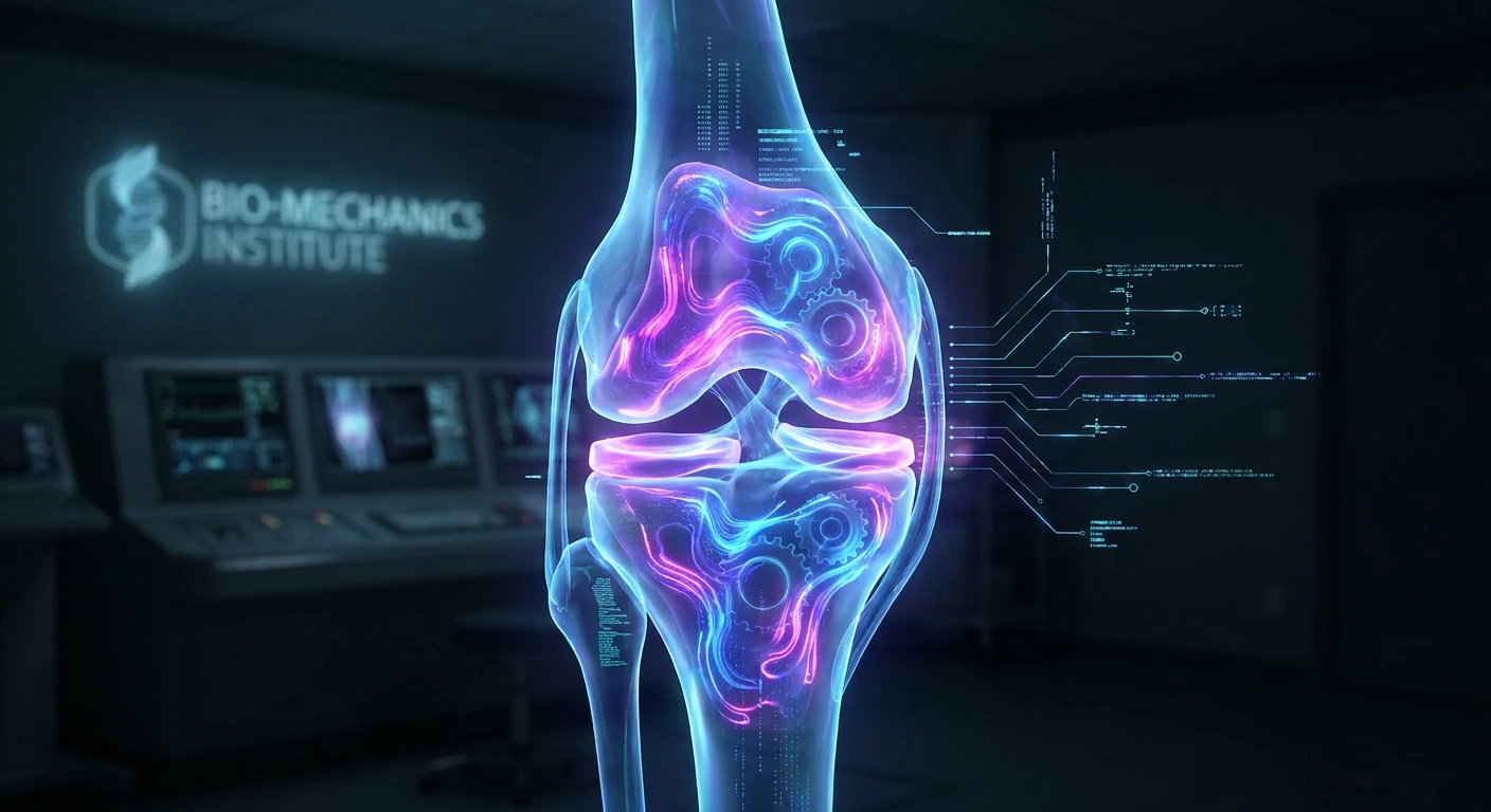
Dealing with sudden or chronic knee pain is always frustrating, especially when it turns out to be a meniscal cyst. A meniscal cyst develops when synovial fluid escapes from the interior of the knee joint, typically through a localized tear in the meniscus cartilage, and pools outside the joint boundaries to form a distinct, fluid-filled sac. You might feel a firm, localized bump right along the joint line of your knee, which is often accompanied by a persistent, dull ache that worsens with physical activity. The meniscus itself serves as a crucial shock absorber, a C-shaped piece of tough, rubbery cartilage that cushions the space between your thigh bone and shin bone. When this cartilage suffers trauma or degenerative wear, it can split. The mechanics of the knee then act like a hydraulic pump. Every time you bend and straighten your leg, you force joint fluid through the tiny tear. This fluid cannot easily return into the joint space, leading to the gradual expansion of the cyst. Understanding the precise anatomical nature of this condition is the first step toward effective management, as treating the swelling alone without addressing the compromised cartilage will inevitably lead to a recurrence of symptoms.
Understanding the Harm, Benefits of Early Detection, and Management
When synovial fluid continues to leak and expand the meniscal cyst, the localized pressure can cause significant harm to surrounding tissues, compressing local nerves and restricting your overall range of motion. If ignored, the underlying meniscal tear will likely propagate, leading to accelerated wear of the smooth articular cartilage and prematurely advancing joint osteoarthritis. Identifying the issue early presents a clear clinical advantage: you gain the opportunity to halt further structural degeneration and preserve the natural biomechanics of your knee. For example, a recreational runner who modifies their training schedule at the first sign of joint line tenderness can often resolve the inflammation through conservative physical therapy. Conversely, an individual who pushes through the pain may eventually require a partial meniscectomy, losing a portion of their natural shock absorber permanently.
Read also: How to Get Rid of a Headache in 2 Minutes Naturally; Lower Back and Thigh Pain Female: Causes and Relief.
| Condition Type | Typical Location | Primary Clinical Signs |
|---|---|---|
| Parameniscal Cyst | Lateral or medial joint line | Palpable firm mass, localized joint line tenderness, pain during deep flexion |
| Baker’s Cyst | Posterior aspect (back of the knee) | Feeling of fullness behind the knee, stiffness, swelling that fluctuates |
| Ganglion Cyst | Various locations near tendons or joints | Usually painless, well-defined margins, translucent under strong light |
Accurate diagnosis requires a structured clinical approach. The following sequential diagnostic steps are standard protocol:
- Comprehensive Physical Examination: A specialist palpates the joint line to locate the firm, localized mass, checking for associated mechanical symptoms like catching, clicking, or locking during knee rotation.
- Musculoskeletal Ultrasound: This non-invasive imaging modality provides immediate confirmation of the fluid-filled sac, easily differentiating it from a solid mass or a benign soft-tissue tumor.
- Magnetic Resonance Imaging (MRI): This is the gold standard diagnostic tool, offering high-resolution visualization of both the cyst and the exact location, size, and geometry of the underlying meniscal tear.
- Diagnostic Arthroscopy: In surgical scenarios, inserting a tiny camera into the joint allows the orthopedic surgeon to directly visualize the cartilage damage and plan the corresponding repair.
Historical Perspectives and the Origins of Knee Pathology
Origins of Knee Pathology Documentation
The medical documentation of localized knee swellings dates back over a century, long before the advent of advanced imaging technologies. Early orthopedic surgeons routinely encountered patients complaining of firm, painful masses along the outer edge of the knee. Without the ability to see inside the joint non-invasively, these masses were frequently misdiagnosed as simple soft tissue tumors, lipomas, or bursitis. Surgical intervention was highly exploratory. Surgeons would perform open arthrotomies—large, invasive incisions—to physically examine the joint space. During these early procedures, medical pioneers began to notice a direct correlation between horizontal cleavage tears of the meniscal cartilage and the presence of these adjacent fluid sacs, establishing the first true anatomical understanding of the parameniscal cyst.
The Evolution of Diagnostic Imaging
The landscape of orthopedic diagnostics shifted dramatically with the introduction of arthrography, a technique where radiographic contrast dye was injected into the knee joint to outline the cartilage structures on an X-ray. While arthrography offered a glimpse into meniscal pathology, it was uncomfortable and lacked precision. The true diagnostic revolution arrived in the 1980s and 1990s with the widespread adoption of Magnetic Resonance Imaging (MRI). MRI provided unparalleled, multi-planar views of soft tissues without exposing patients to ionizing radiation. Radiologists could finally trace the microscopic pathway of fluid escaping from the central joint cavity, through the damaged meniscal fibers, and pooling into the surrounding parameniscal tissues. This technological leap eliminated diagnostic guesswork, allowing surgeons to precisely map the lesion before the patient ever entered the operating room.
Modern State of Arthroscopic Surgery
Today, the management of a meniscal cyst is highly refined, heavily favoring minimally invasive techniques. Modern arthroscopic surgery utilizes high-definition fiber-optic cameras and precision micro-instruments that require incisions no larger than a keyhole. Instead of aggressive, open joint procedures that necessitate prolonged hospital stays, patients now typically undergo outpatient arthroscopy. The modern surgical philosophy prioritizes cartilage preservation. Surgeons aim to meticulously repair the tear using specialized sutures or, if repair is impossible, perform a highly selective partial meniscectomy to remove only the damaged, non-functional tissue. This evolution reflects a broader medical shift toward minimizing surgical trauma, optimizing rapid functional recovery, and actively preventing long-term degenerative joint disease.
The Pathophysiology and Biomechanics of Synovial Extravasation
The Pathophysiology of Synovial Fluid Extravasation
The fundamental mechanism driving the formation of a meniscal cyst relies on the unique hydraulic properties of the human knee joint. Synovial fluid is a viscous, lubricating substance produced by the synovial membrane to reduce friction and supply nutrients to the avascular cartilage. When a horizontal cleavage tear develops in the meniscus, it creates a microscopic channel communicating from the central articular cavity to the periphery of the knee. As you stand, walk, or run, the immense compressive forces generated within the joint physically squeeze the synovial fluid out through this tear. Because the torn edges of the cartilage often act as a mechanical one-way valve, the expelled fluid cannot flow backward into the joint space when the pressure is released. Over weeks or months, the constant, unidirectional pumping action forces the fluid to accumulate within the parameniscal soft tissues, encapsulating itself in a fibrous sac.
Biomechanical Impact on the Knee Joint
The presence of a localized fluid collection significantly alters the normal biomechanics of the knee. The knee joint relies on a delicate balance of tension between ligaments, tendons, and cartilage to maintain stability during complex movements. A firm cyst sitting on the joint line acts as a mechanical obstruction. During extreme ranges of motion, particularly deep flexion or forced rotation, the surrounding soft tissues are unnaturally stretched over the expanded sac, generating acute, localized pain. Furthermore, the underlying meniscal deficiency means that the articular surfaces of the femur and tibia lose their primary shock absorber in that specific compartment, increasing localized contact stress and accelerating cellular degradation.
- Synovial Fluid Composition: The fluid trapped within the sac is highly rich in hyaluronic acid, mucin, and lubricin, giving it a thick, gelatinous consistency similar to a heavy lubricating oil.
- Lateral vs. Medial Prevalence: Statistical analyses consistently demonstrate that these cystic formations are significantly more common on the lateral (outer) side of the knee compared to the medial (inner) side, largely due to the differing anatomy and mobility of the lateral meniscus.
- Associated Cartilage Lesions: Over 85 percent of all parameniscal swellings are directly associated with an underlying horizontal cleavage tear, emphasizing that the mass is a secondary symptom rather than an isolated primary disease.
- Capsular Distension: The chronic accumulation of fluid leads to a progressive, microscopic distension of the joint capsule, which triggers local nociceptors and causes the deep, aching sensation reported by most patients.
A Comprehensive Step-by-Step Action Plan
Step 1: Immediate Activity Modification
The moment you identify a hard, painful lump on your knee joint line, you must immediately halt all high-impact activities. Continuing to run, jump, or perform heavy squats will only increase the intra-articular pressure, forcing more fluid into the sac. Transition exclusively to low-impact exercises, such as swimming or upper-body strength training, to maintain your cardiovascular fitness without stressing the compromised meniscal tissue.
Step 2: Cryotherapy and Compression
Managing acute inflammation is critical in the early stages. Apply ice packs to the affected area for twenty minutes every few hours, ensuring a physical barrier like a towel sits between the ice and your skin to prevent frostbite. Utilizing a high-quality elastic compression sleeve can also help mitigate generalized knee swelling, though it will not physically reduce the internal volume of the established fluid sac.
Step 3: Medical Evaluation and MRI
Do not attempt to self-diagnose joint pathology. Schedule an appointment with an orthopedic specialist who will perform a manual examination to assess joint stability and pinpoint the exact source of tenderness. The physician will order a high-resolution MRI to map the internal architecture of your knee, confirming the exact size of the swelling and the precise geometry of the associated horizontal cleavage tear.
Step 4: Non-Surgical Interventions
If the symptoms are mild and the underlying tear is small, your doctor may recommend a conservative approach. This often involves a short course of non-steroidal anti-inflammatory drugs (NSAIDs) to reduce systemic inflammation. In certain cases, an ultrasound-guided corticosteroid injection may be administered directly into the cyst or the adjacent joint space to rapidly suppress local tissue inflammation and alleviate pain.
Step 5: Pre-Operative Physical Therapy
Whether you eventually require surgery or not, engaging in targeted physical therapy is essential. A specialized physical therapist will guide you through strict, non-weight-bearing isometric exercises designed to strengthen the quadriceps, hamstrings, and gluteal muscles. Building robust muscular support around the knee joint reduces the direct mechanical load transferred through the damaged meniscal cartilage during daily activities.
Step 6: Arthroscopic Decompression
If conservative treatments fail to provide lasting relief, minimally invasive arthroscopic surgery becomes necessary. The surgeon makes two tiny incisions, inserting a high-definition camera and precision micro-shavers. The primary goal is to identify the one-way valve effect caused by the torn meniscus. By carefully trimming away the frayed cartilage edges or suturing the tear closed, the surgeon effectively stops the hydraulic pump mechanism, naturally decompressing the localized fluid collection.
Step 7: Post-Surgical Rehabilitation
Surgical intervention must be immediately followed by a structured rehabilitation protocol. The initial weeks focus on gentle range-of-motion exercises to prevent the formation of internal scar tissue and eliminate joint stiffness. As healing progresses, the protocol shifts toward progressive resistance training and proprioceptive balance drills. Most patients can expect to safely return to full, unrestricted athletic activities within four to eight weeks, provided they adhere strictly to the therapist’s guidelines.
Myths and Reality
Myth: A meniscal cyst is a dangerous, malignant tumor that requires aggressive oncology treatment.
Reality: These swellings are entirely benign, non-cancerous collections of naturally occurring synovial fluid that pose no systemic threat to your health.
Myth: You can permanently cure the bump by simply having a doctor drain it with a syringe.
Reality: Needle aspiration only addresses the superficial symptom. Because the underlying cartilage tear continues to act as a one-way pump, the fluid will almost certainly reaccumulate shortly after being drained.
Myth: Only elite, high-performance athletes experience this specific type of joint condition.
Reality: While trauma can cause it, routine joint degeneration and mild osteoarthritis can easily create microscopic meniscal tears in older adults, leading directly to fluid accumulation regardless of athletic background.
Myth: Developing this condition means you will inevitably need a total knee replacement.
Reality: The vast majority of cases resolve perfectly through conservative physical therapy or a minor, highly successful outpatient arthroscopic procedure to repair the torn cartilage.
Frequently Asked Questions and Conclusion
Does a meniscal cyst ever heal completely on its own?
While the visible swelling may fluctuate in size depending on your daily physical activity level, the underlying structural cartilage tear rarely heals spontaneously without targeted clinical intervention, biomechanical correction, or significant rest.
Is stationary cycling considered a safe exercise for this condition?
Cycling is generally categorized as a low-impact and safe alternative, provided you keep the mechanical resistance very light and ensure the bicycle seat height is perfectly adjusted to prevent excessive, loaded knee flexion.
Can wearing a tight compression brace actually help?
A high-quality knee sleeve can provide temporary symptomatic relief from the generalized aching sensation and help manage overall joint swelling, but it cannot exert enough pressure to eliminate the localized, internal fluid sac.
What physically happens if the fluid sac bursts?
Though quite uncommon, a spontaneous rupture can cause a sudden, sharp localized pain followed by a feeling of mild warmth as the viscous fluid disperses into the surrounding soft tissues, where the body will gradually and naturally reabsorb it.
Can a standard musculoskeletal ultrasound accurately detect the swelling?
Yes, diagnostic ultrasound is an excellent, quick, and completely non-invasive imaging tool to reliably confirm the presence of a fluid collection right at the joint line, though it is less effective than MRI at visualizing the deep cartilage tear.
How long is the typical functional recovery after arthroscopy?
Most patients can walk comfortably without crutches within a few days and safely return to non-impact sports or rigorous daily activities within four to six weeks following an uncomplicated arthroscopic decompression and partial meniscectomy.
Is it structurally possible for the swelling to return after surgery?
Recurrence is statistically highly unusual if the operating surgeon successfully identifies, addresses, and fully resolves the underlying one-way valve tear during the initial arthroscopic procedure.
What exactly occurs if I completely ignore the chronic discomfort?
Ignoring the persistent mechanical symptoms can lead to the meniscal tear expanding further, which rapidly accelerates the breakdown of the surrounding articular cartilage and significantly hastens the early onset of debilitating osteoarthritis.
Navigating the complexities of joint pain requires patience, accurate information, and a proactive mindset. A meniscal cyst is a clear biological signal that your knee’s internal mechanics are compromised. By understanding the direct connection between cartilage damage and synovial fluid accumulation, you can make informed decisions about your rehabilitation. Whether you opt for conservative management or minimally invasive arthroscopy, prioritizing the long-term health of your joint is paramount. Do not let chronic pain dictate your active lifestyle—consult with a certified orthopedic specialist today, map out a definitive diagnostic plan, and take the first decisive step toward restoring your knee’s natural, pain-free mobility.




Leave a Reply