

Understanding hpv bumps on back of tongue: What You Need to Know
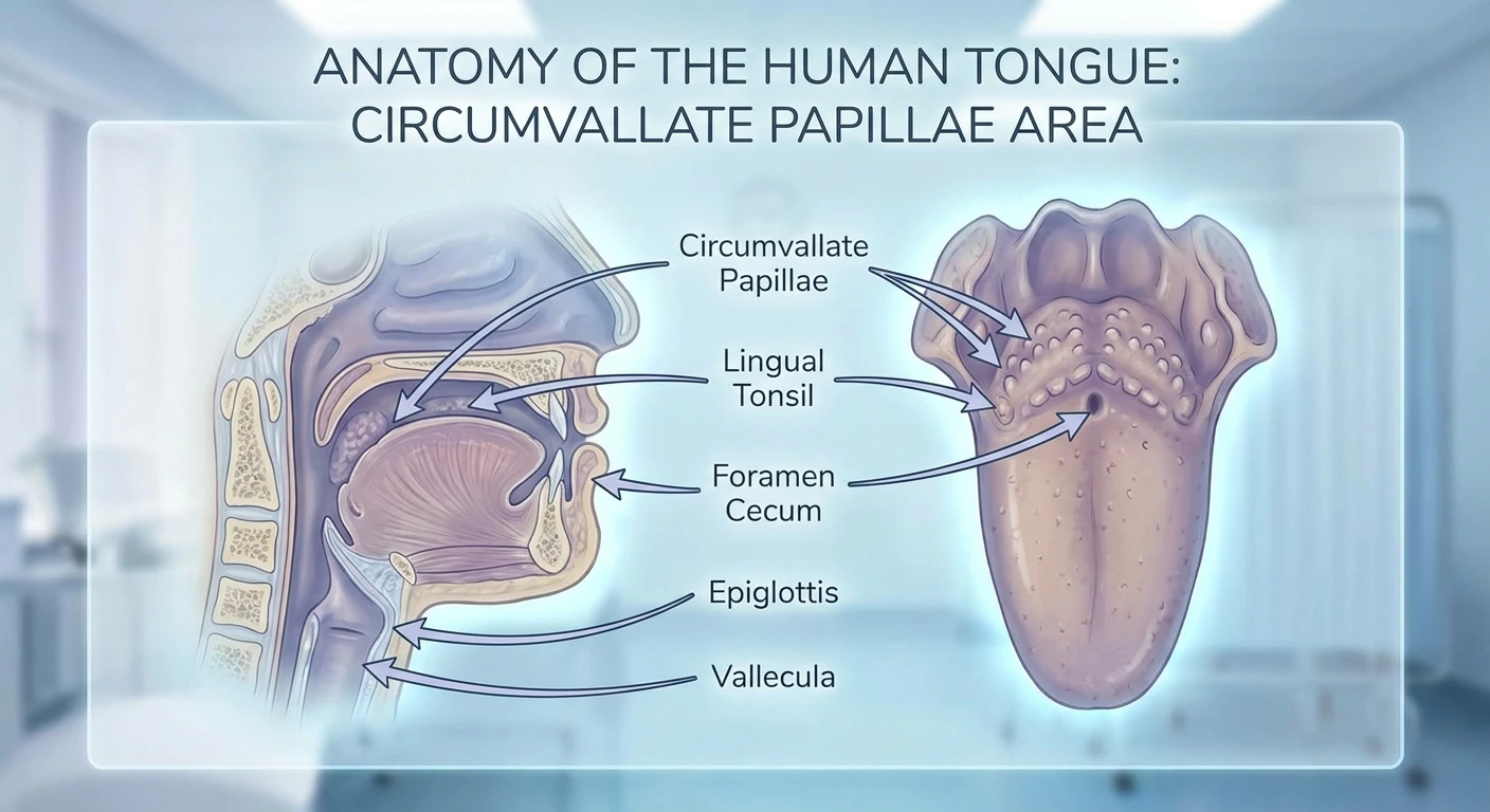
If you recently noticed unusual textures or growths in your mouth, you might be urgently searching for information on hpv bumps on back of tongue. Let us clear the air right away: finding strange bumps in the mirror can trigger a lot of immediate anxiety, but distinguishing between completely normal anatomical structures and actual viral infections is your first line of defense. Many people look closely at their tongue for the first time, spot the large, naturally occurring circumvallate papillae at the very back, and immediately assume they have contracted a viral disease. However, the Human Papillomavirus can indeed cause genuine lesions in the oral cavity. Understanding the difference between normal anatomy and viral infections saves you unnecessary anxiety while ensuring you get the exact right medical care if you actually have an active oral infection.
This detailed guide delivers clear, factual context about how oral tissues behave, how a viral infection physically manifests on mucous membranes, and what exact steps you should take next. You will gain a thorough understanding of the clinical signs of oral lesions, ensuring you are equipped with medical knowledge rather than internet-fueled panic.
Read also: Fix White Spots On Tongue: Causes & Tips; Symptoms of Montgomery Gland Cancer Explained.
Why Early Differentiation Matters
Accurately identifying the nature of oral bumps offers immense benefits. First, it prevents the psychological harm of misdiagnosing your own natural taste buds as a sexually transmitted infection. Second, if you truly do have a viral papilloma, catching it early prevents it from growing larger or spreading to adjacent tissues. A clear value proposition emerges when you monitor your oral health proactively: you catch benign oral warts while they are easily treatable, and you maintain vigilant screening for high-risk strains that could eventually lead to oropharyngeal complications.
For example, if an individual notices a small, cauliflower-like growth and visits an Ear, Nose, and Throat (ENT) specialist immediately, a simple, painless excision can resolve the issue within minutes. Conversely, another individual might ignore a continuously growing, persistent lesion for months, requiring much more invasive biopsy procedures later. Early action always favors your overall health.
| Feature | Normal Circumvallate Papillae | Viral HPV Lesions |
|---|---|---|
| General Appearance | Smooth, round, uniform, and symmetrical bumps arranged in a V-shape. | Irregular, rough, sometimes cauliflower-like or spiky (papillary) surface. |
| Location | Strictly at the very back of the tongue, forming a distinct horizontal line. | Can appear anywhere: back of tongue, roof of mouth, inner cheeks, or lips. |
| Color and Sensation | Pinkish, matching the rest of the tongue. Usually painless unless burned by hot food. | Often white, pink, or slightly red. Painless, but may feel like a foreign object. |
If you spot suspicious growths that do not match standard tongue anatomy, follow these exact steps to protect your health:
- Monitor the duration of the bump; natural irritation usually heals within 14 days, whereas viral lesions persist.
- Document any changes in size, color, or texture using your smartphone camera with the flash turned on.
- Book an appointment with a qualified dentist, oral surgeon, or ENT specialist rather than attempting a home remedy.
- Avoid aggressive brushing or scraping of the area, which can cause bleeding or secondary bacterial infections.
Origins of Oral HPV Discoveries
The journey to understanding how certain viruses interact with oral tissues has a rich medical history. Decades ago, clinicians often struggled to correctly classify unusual growths in the mouth. Before the advent of advanced molecular virology, many benign oral warts were frequently misdiagnosed as signs of other systemic infections, such as syphilis, or merely dismissed as localized traumatic ulcers caused by biting or friction. It was not until the late 20th century that scientists definitively mapped the genetic structures of the Human Papillomavirus, realizing that the same family of viruses responsible for cutaneous skin warts and genital warts could also colonize the mucosal lining of the mouth and throat.
Evolution of Viral Strains
As DNA sequencing technology advanced, researchers began categorizing the virus into distinct numerical strains. They discovered that low-risk strains, specifically types 6 and 11, are the primary culprits behind benign squamous cell papillomas—the non-cancerous, typical warts found on the tongue and palate. These strains evolved to aggressively hijack the basal cells of the epithelium, forcing rapid but localized cellular replication that creates the physical bump. Meanwhile, high-risk strains, such as type 16, were identified as having a completely different evolutionary mechanism, often integrating directly into the host DNA and bypassing the creation of a physical wart in favor of long-term cellular mutation.
Modern State of Oral Health Screenings
Today, the medical community’s approach to oral lesions is highly sophisticated. Routine dental exams now frequently include comprehensive oral cancer and viral lesion screenings as standard practice. Dentists are trained to use specialized fluorescent lights and distinct visual criteria to differentiate between normal lingual tonsils, everyday canker sores, and true papillomas. Furthermore, widespread vaccination campaigns introduced in the 21st century have dramatically altered the landscape, significantly reducing the incidence of new infections among vaccinated populations and providing robust protection against both the benign bump-causing strains and the severe, cancer-causing variants.
The Cellular Mechanics of Papillomavirus
To truly understand how these growths form, you have to look at the microscopic cellular mechanics of the human mouth. The mouth is lined with something called stratified squamous epithelium. This is a multi-layered protective barrier of flat cells designed to withstand the daily friction of chewing, swallowing, and speaking. For the virus to establish an infection, it must bypass the tough upper layers of this barrier. It typically does this through microscopic tears or micro-abrasions in the tissue—often caused by something as simple as eating hard foods, accidental cheek biting, or routine dental work.
Understanding Squamous Epithelium
Once the virus reaches the deepest layer, known as the basal layer, it enters the stem cells. Instead of immediately destroying the cell, the virus inserts its genetic material and essentially reprograms the cell’s internal machinery. As these infected cells divide and push upward toward the surface of the tongue, they do not mature and flatten out normally. Instead, they multiply rapidly and chaotically, creating a structural buildup of excess tissue. This localized cellular traffic jam is what eventually becomes visible to the naked eye as a textured bump.
- The virus causing these lesions is a non-enveloped, double-stranded DNA virus, making it highly resilient in warm, moist environments.
- Over 200 distinct strains of this virus exist, with approximately 40 specifically adapted to thrive in mucosal environments like the mouth and throat.
- The physical bumps caused by low-risk strains are clinically referred to as squamous cell papillomas, condyloma acuminatum, or verruca vulgaris.
- The human immune system is incredibly efficient; clinical data shows that macrophages and T-cells naturally clear over 90 percent of these mucosal infections within two years without any medical intervention.
Day 1: Initial Visual Inspection
Begin your proactive health check by securing a bright flashlight and a clean mirror. Wash your hands thoroughly. Open your mouth wide and gently extend your tongue. Look specifically at the very back. You will naturally see a V-shaped row of large bumps—these are your circumvallate papillae and they belong there. Note if there are any isolated, irregular, or cauliflower-shaped growths separate from this symmetrical V-shape. Document the baseline appearance.
Day 2: Tracking Physical Sensations
Focus entirely on sensory feedback. Do you feel a persistent lump when you swallow? Is there any pain when consuming acidic foods like citrus or tomatoes? Viral papillomas are overwhelmingly painless, acting merely as physical obstructions. If the bump is highly painful, burns, or stings, it is much more likely to be a canker sore (aphthous ulcer) or a traumatized taste bud rather than a viral wart. Write down your observations.
Day 3: Oral Hygiene Optimization
Evaluate and adjust your oral care routine. Switch immediately to a soft-bristled toothbrush to prevent further micro-abrasions to your oral mucosa. Discontinue the use of harsh, alcohol-based mouthwashes, as these can dry out the tissues and cause further cellular irritation. Stick to gentle flossing and mild fluoride toothpaste. Creating a calm, non-irritated environment helps you see if the bump resolves on its own due to reduced friction.
Day 4: Logging Lifestyle Factors
Take inventory of your daily habits. Are you a heavy smoker or frequent consumer of alcohol? Both of these factors severely dry out the oral mucosa and suppress local immune responses, making it harder for your body to fight off mucosal infections. Additionally, note if you have a habit of chewing on the inside of your cheeks or biting your tongue, as trauma creates the exact entry points the virus exploits.
Day 5: The Lymph Node Check
Perform a gentle physical palpation of your neck. Use the pads of your fingers to feel along the sides of your neck, just under your jawline, and behind your ears. You are checking for swollen, hard, or tender lymph nodes. While benign viral bumps rarely cause lymph node swelling, persistent swelling combined with oral lesions is a clinical signal that warrants immediate professional medical evaluation to rule out more serious pathology.
Day 6: Booking a Professional Evaluation
If the bump has remained unchanged or grown over the past six days, it is time to schedule an appointment. Contact your dentist, an oral surgeon, or an ENT doctor. When you call the front desk, be specific: state that you have a persistent, painless growth on the back of your tongue that you would like evaluated for a potential biopsy or removal. Specialists handle these routine checks daily.
Day 7: Preparing Your Medical History
Gather all relevant information before your appointment. Write down exactly when you first noticed the bump, any changes in its size or texture, and a list of any current medications you are taking. Be prepared to discuss your vaccination history openly. Providing your doctor with a clear, factual timeline eliminates guesswork and ensures you receive the most accurate diagnostic procedure, whether that is simple monitoring, freezing, or surgical excision.
Myth: Every bump at the back of the tongue is a viral infection.
Reality: The vast majority of people who panic over bumps at the back of their tongue have merely discovered their circumvallate papillae or lingual tonsils. These are normal, natural anatomical structures involved in tasting and immune function, completely unrelated to any virus.
Myth: Oral viral lesions always develop into severe mouth cancer.
Reality: The strains that cause visible, cauliflower-like warts (strains 6 and 11) are considered low-risk and are practically never associated with cancer. The high-risk strains linked to cancer typically do not produce visible warts at all.
Myth: You can cure viral mouth bumps by gargling strong mouthwash.
Reality: No over-the-counter mouthwash, essential oil, or home remedy can cure a cellular viral infection. The immune system must clear the virus naturally, or a doctor must physically remove the lesion.
Myth: Only people with poor hygiene develop these oral growths.
Reality: This virus is the most common infection of its kind globally. Almost all sexually active adults will encounter at least one strain in their lifetime, regardless of their oral hygiene habits.
Are normal tongue bumps contagious?
No. Normal anatomical bumps like circumvallate papillae or inflamed taste buds are part of your body and cannot be passed to anyone else.
Can a dentist diagnose oral warts?
Yes. Dentists are highly trained in oral pathology and are often the first medical professionals to identify, diagnose, and manage abnormal oral growths.
Do these viral lesions bleed easily?
Generally, benign viral papillomas do not bleed unless they are aggressively bitten, scratched, or traumatized by hard foods and toothbrush bristles.
How long do oral warts last without treatment?
While the immune system clears most infections within one to two years, a visible oral wart may persist for months and usually requires minor physical removal by a doctor.
Is there a vaccine for mouth infections?
Yes. The widely available recombinant vaccines protect against the most common high-risk and low-risk strains, significantly reducing the chances of developing oral lesions.
Can I scrape off an oral papilloma at home?
Absolutely not. Scraping or cutting an oral lesion at home can lead to severe bleeding, intense bacterial infection, and the further spread of the virus to other areas.
Does smoking make these lesions worse?
Yes. Smoking suppresses local oral immunity, alters the protective mucosal barrier, and increases the risk of viral persistence and cellular mutation.
Conclusion
Finding unexpected textures in your mouth can be alarming, but maintaining a factual, calm perspective is essential. Most bumps on the back of the tongue are entirely natural anatomical features that have always been there. However, if you genuinely suspect you have persistent viral lesions, modern medicine offers incredibly simple, highly effective solutions. Do not rely on self-diagnosis or internet speculation. Take control of your oral health, monitor the area strictly for changes, and schedule a professional medical checkup today to secure peace of mind and excellent long-term health.





Leave a Reply